
OPTICAL ILLUSIONS
By Judy Quinones, ABO-AC, NCLE-AC
Learning Objectives:
Upon completion of this program, the participant will be able to:
- Explain how our brain processes visual stimuli.
- Explain the difference in the role of the human eyes versus the human brain in seeing optical illusions.
- Appreciate the complexity of the human visual system.
- Classify the differences between the three main categories of optical illusions.
Faculty/Editorial Board
 Judy graduated summa cum laude from the Ophthalmic program at Raritan Valley Community College, where she won the Excellence in Ophthalmics award for the class of 2022.
She won the College Bowl at Vision Expo East held in New York City the same year.
Judy graduated summa cum laude from the Ophthalmic program at Raritan Valley Community College, where she won the Excellence in Ophthalmics award for the class of 2022.
She won the College Bowl at Vision Expo East held in New York City the same year.
Judy is a Licensed Dispensing Optician in the State of NEw Jersey, and she holds advanced ABO and NCLE certifications.
Judy is an Advanced Ophthalmic Speaker. Prior to embarking on her path within opticianry, she earned a degree in Economics with magna cum laude honors.
Among her roles, Judy enjoys her role as a preferred substitute teacher. Fluent in multiple languages she enjoys traveling, fine food, and cultural learning. But, the role she cherishes most is that of a loving dedicated mother.
Credit Statement:
This course has been approved for one (1) hour of CE credit by the American Board of Opticianry - ABO, Ophthalmic Technical Level 3. Course number: STWJHI084-3
What you see is not always what you get! If you want to be optically amazed, this course is for
you. In this course, we explore the cause of optical illusions and why the brain is tricked into perceiving what
is not there. The aim of this course it to help you understand
how optical illusions can deceive your brain and give insight
into the visual system’s complexity.
Optical Illusions are fascinating and mind boggling at the same time. Some refer to optical illusions as a trick of the eye.
An optical illusion is defined as a misleading image presented to the vision (Definition of Optical Illusion, 2019). How is it
possible to see something that is not real? When we understand that we do not see with our eyes, but instead with our
brains and that the process is a very complex, then optical illusions and their trickery make more sense. While most of us
find optical illusions entertaining, medical and scientific researchers find them valuable tools that can reveal the hidden
constraints of the perceptual system in a way that normal perception does not (Seckel, 2000).
Anna Claybourne (2020) explains how our eyes and brain are constantly flooded with images all day. Our brain, tasked
with making sense of these images in split seconds, takes shortcuts based on memories of previous experiences, decides what
information is relevant, fills in the blanks to save us time and produces what we are seeing. A simplified example of our
brain filling in the blanks for us is apparent when reading the following text found in Brown & Bornoff (2017):
It deosn’t mttaer in what order the ltteers in a word are, the only iprmoetnt thing is that the frist and lsat ltteer be at
the rghit pclae. This is bcuseae the human mind deos not raed ervey lteter by istlef, but the word as a wlohe.
HISTORY OF OPTICAL ILLUSIONS
The discussion and debate of optical illusions date back to ancient Greece, where they were used in architecture and art. Greek philosophers such as Epicharmus, Protagoras, Plato and Aristotle tried to explain how our eyes, our brain, or both, trick us into seeing things that are not there. In the 19th century, psychologists Johannes Mueller and J.J. Oppel revived the interest in visual illusions. They conducted many studies that led to several articles published on the topic. German physicist Herman Von Helmholtz, also in the 19th century, stated that illusions happen when our preconceived notions of reality do not match what we see, calling it a cognitive illusion. From here, cognitive illusions were divided into four types: distorting, paradox, fiction and ambiguous (Optical Illusions: A Brief History, 2021). As we can see, optical illusions have been of interest to ancient Greek philosophers and the many physicists, sociologists, psychologists, artists, vision scientists and others seeking explanations for these visual deceptions.
PATHWAY TO VISION
How do we see? One way of answering this question is by tracing a light ray from an object we are looking at and following its path. Light reflects or bounces off an object, and that light ray enters our eye, traveling through the tear film, cornea, aqueous, pupil, more aqueous, lens, vitreous and finally hitting the retina. The retina is an extension of the brain, and we see with our brain, therefore the study of optical illusions lies with a second, more complicated pathway starting in the retina and ending in the brain’s visual cortex (Fig. 1). Each of our two retinae are lined with light-sensitive photoreceptor cells called cones and rods. The cone cells are used for photopic (daylight) vision and produce color vision. They are highly concentrated in the fovea of the macula, at the center of the retina. The fovea is the area of the retina that produces the highest resolution, color vision and detail.
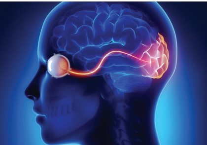
FIG. 1. The visual cortex of the brain is where optical or visual illusions form; the brain will try and fail to correctly interpret images from patterns, light and color. The deceptive images perceived are what we call optical illusions
Cone photoreceptors are commonly classified into three types depending on their peak response to wavelengths of red, green or blue light. Rod cells, on the other hand produce no
color vision, only shades of gray and are found mainly in the periphery of the retina. Rod photoreceptor cells are responsible for our scotopic or dark-adapted vision in dim-low
illumination levels. Our retinal photoreceptors cells convert light into electrical impulses. These electrochemical signals travel to the brain along our two optic
nerves (Molday & Moritz, 2015).

The optic nerve, which consists of about two million nerve fibers, transmits the visual information from each retina. These
nerves cross over at the optic chiasm, allowing our visual cortex to obtain information from both of our eyes (Fig. 2). This process allows the images from both
eyes to generate depth perception. The information continues along the optic tracts to the thalamus via the LGN (lateral geniculate nucleus) and the superior colliculus.
The superior colliculus moves the eyes in short jumps, called saccades. Saccades allow the brain to perceive a smooth scan by stitching together a series of
relatively still images (Daw, 2012). This visual information continues to be transmitted through the optic radiations onto the visual cortex, where the images
captured by the retina begin to get processed and recognized by our brain. It is within the six layers of the visual cortex where depth perception, form, color and
motion are perceived (The visual pathway from the eye to the brain, 2021). In the brain’s occipital cortex, electrical impulses from the retina are interpreted and
perceived as images and inverted retinal images are flipped to appear right side up. The visual cortex is divided into six critical areas, which are arranged hierarchically depending on the structure and function of
the area. These are often called V1, V2, V3, V4, V5 and the inferotemporal cortex. The primary visual cortex (V1) is the first stop for visual information in
the occipital lobe (Guy-Evans, 2021). From here, the visual cortex filters the visual information. It sends it to other areas of the brain responsible for more complicated visual tasks (Common Causes of Visual Loss in
Children, n.d.).
The visual cortex is capable of visual tasks including perceiving the position of objects, details, patterns and
coordinating the information from each eye, known as binocular vision. However, more complicated visual tasks such as color vision analysis and detailed
motion processing occur in areas of the brain that are dedicated to this purpose and others. The flow of
information is organized from here on so that the correct signals reach the right parts of the brain. Our brain’s high-level system is so sophisticated and unique
that the mechanisms to understand how it all works are not yet fully understood. The fact that the visual system cannot be replicated using even the highest
spec computer is a testament to the super-advanced processing capabilities which exist within the human brain (Woodruff, 2017)
OPTICAL ILLUSIONS AND THE BRAIN'S RESPONSE TO THEM
At what point in the visual pathway from the retina to the many cortical visual areas does the neural activity
correlate with what we perceive? Do neurons in the retina, primary visual cortex (V1) and higher-level cortical areas contribute to perception equally? Or
instead, does perception have a specific locus in the brain (Shimojo et al., 2001)? Questions like these remain largely unanswered. Scientists are still unable
to figure out where and how, along the path from the eyes to the cortex, the sensory input perceived by the
retina is converted into a meaningful object representation, which can be consciously manipulated by the brain (Diamant, 2008).
Over 50 percent of the cortex, the brain’s surface, is used to process visual information. This is why it is believed that understanding how vision works will
also be a key piece in understanding how the brain as a whole works. While software has been developed to tackle many challenges, computer models barely
scratch the surface of human vision. Human vision has been compared to a camera in those images we see are cast on the retina and sent to the brain. But David
Knill, a professor of brain and cognitive sciences at the University of Rochester, says that human vision is more akin to speech than photography. From the time
we are infants, our brain is learning how to build a three-dimensional environment through the interpretation of visual sensory signals such as shape, size and
occlusion. We basically learn to see, and our sight is constantly adapting (Rochester Review: University of Rochester, n.d.).
A team of neuroscientists at MIT found that the human brain is capable of processing images that it sees for as little as 13 milliseconds. Through their
study, they concluded that vision involves finding concepts and that the human brain tries to understand what it is looking at all day long. This same study
briefly describes what happens to the visual input after it reaches the retina. This information flows to the brain, where information that has to do with shape,
color and orientation is further processed. Past studies suggested that the flow of visual information went from the retina to the “top” of the visual processing
chain in the brain and back down again for further processing. This study, however, offered evidence that
“feedforward processing,” meaning the flow of information in only one direction, from the retina through
visual processing centers in the brain, is enough for the brain to identify concepts without having to do any further feedback processing (In the blink of an eye, n.d.).
The human brain is capable of detecting and classifying everyday objects, such as a coffee cup, words on a page, etc.,
from among tens of thousands of possibilities within a fraction of a second, despite the enormous variation in appearance that each of these items
produces on our eyes. From an evolutionary perspective, these recognition abilities are expected. Humans need to be able to accurately and quickly identify
objects that are projected to the photons on our retinae. However, even today, there is no clear and undisputed answer to how our visual system and brain solve
object recognition: How does it go from taking each retinal image, and identifying and categorizing these objects? Neuroscientists are interested in mapping the
spatial layout and connectivity of the relevant brain areas, uncovering conceptual definitions, and reaching
cellular and molecular targets that can be used to predictably modify object perception. For example, by uncovering the neuronal circuitry underlying object
recognition, we might ultimately repair that circuitry in brain disorders that impact our perceptual systems, such as blindness or agnosias (DiCarlo et al., 2012).
Neuropsychologist Richard Gregory, in his book Seeing Through Illusions, expresses that we can only believe what we see with our eyes up to a certain point,
for we are constantly deceived. Why are we deceived? It is because we rely on clues like shading and color strength for depth, and on a series of assumptions of
how our world functions. So, in short, it does not come down to what we see as much as how we interpret what we are seeing. We expect that an object at a
distance will be smaller than one up close and the same size; however, when these basic assumptions are tested, we tend to fail. Our memory bank is full of
past experiences and populated by what we have learned to see (Gregory, 2009).
Visual illusions stem from a perception that differs somehow from the physical stimulus. As such, visual illusions are powerful non-invasive tools that can
help us better understand the process by which the brain perceives and processes visual stimuli (Gori et al., 2016).
Our visual system is tasked with receiving not only two slightly differently positioned images from each of our retinas, referred to as binocular disparity but
also two- dimensional images from a three-dimensional world. This binocular disparity helps the brain figure out the distance. Still, there is also the question
of lightness and depth perception, which play into the relationship between brain activity and conscious visual perception. Research has determined that visual
perception must involve progressive computations in multiple areas of the brain and hierarchies of visual computations (Shimojo et al., 2001). Added to this is
that the brain must process this information very quickly and reach one decision. And it is here where the brain must resort to prior knowledge.
The main task of human perception is to amplify and strengthen sensory inputs to be able to perceive, orientate, and act very quickly, specifically and efficiently (Carbon, 2014).
CLASSIFICATION OF OPTICAL ILLUSIONS
In his book, The Ultimate Book of Optical Illusions, Seckel (2006) lists 349 of what he refers to as the world’s most powerful optical illusions. Many
attempts have been made at classifying the vast number of optical illusions with three types of illusions
being generally recognized: literal illusions, physiological illusions and cognitive illusions.
“Literal optical illusions are two different images compressed into one. Physiological optical illusions overstimulate the brain with light, shapes
and colors. Cognitive optical illusions are like logical paradoxes that present inherently contradictory information.” (bigthink.com/neuropsych/optical-illusions-art-science)
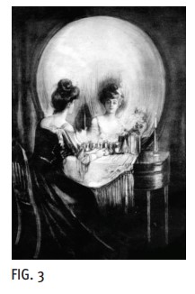
Literal illusions are typically produced by assembling multiple images to confuse the viewer. One can literally see two different images in the same
picture depending on the viewers perspective. Each component may be easy to view, but when seen as a whole, they may look vastly different from the parts. This type of
illusion is based on the phenomenon known as filling-in. In Fig. 3, a woman is seen in front of a mirror, and alternately the mirror and vanity table
appear as a skeleton. Both images exist so the final result you see in a literal illusion depends on your perception.
Physiological illusions: When eyes are overstimulated, shapes and colors appear that aren’t there.
An example is the Hermann Grid illusion in Fig. 7. These are commonly characterized by the afterimages after looking at bright lights. This illusion is
formed by a repetitive or intense stimulus that leads us to perceive false movement or repetition. Another example of an after-image can be seen in Fig. 4 when we switch to look at the small cross in the
white square on the right after staring at the white cross in the middle of the blue cross for 30 seconds.
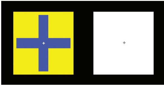
FIG. 4 Physiological illusion: Look at the + in the middle of the blue figure above for 30 seconds. Then look at the tiny + in the center of the white square on the right.
Cognitive illusions occur when the brain makes erroneous assumptions about an object; an example of an ambiguous illusion is the Necker Cube. It uses a wireframe cube named after the Swiss crystallographer Louis Necker (1880s) who observed that cubic shapes repeatedly reverse their perceived orientation. In Fig. 5, the stairs reverse direction if you stare at them for roughly 30 seconds. Cognitive illusions result from our brain’s conceptions and assumptions about the world, which we impose upon visual stimuli.
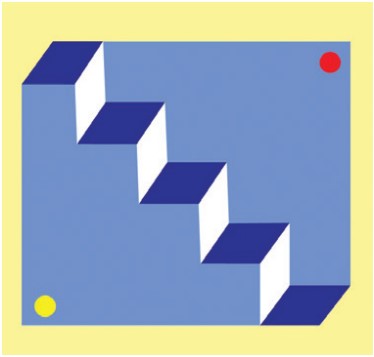
FIG. 5 Ambiguous illusion, Schroeder’s Stair: Stare at the stairs until they reverse direction.
SOME FAMOUS ILLUSIONS EXPLAINED
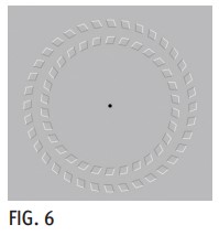 The Pinna-Brelstaff Revolving Circles Illusion (Fig. 6) is named after the Italian vision scientist B. Pinna, who discovered it in
1990 and an English psychologist, G. Brelstaff. This motion illusion is the first to show a rotating motion phenomenon. It comprises concentric
rings consisting of small diamond-shaped figures angled in different directions for each ring. After staring at the center of the image, move your head toward
the image, and the circles appear to turn clockwise. Then, as you move your head away from the image, the circles counter-rotate. The movement that we see is achieved by the angle
the shapes. No one is exactly sure why this happens, but a theory is that there is a communication delay between the regions of the brain that process
vision (Pinna, 2009). he Hermann Grid Illusion (Fig. 7) was created by German physiologist Ludimar Hermann in 1870.
The Pinna-Brelstaff Revolving Circles Illusion (Fig. 6) is named after the Italian vision scientist B. Pinna, who discovered it in
1990 and an English psychologist, G. Brelstaff. This motion illusion is the first to show a rotating motion phenomenon. It comprises concentric
rings consisting of small diamond-shaped figures angled in different directions for each ring. After staring at the center of the image, move your head toward
the image, and the circles appear to turn clockwise. Then, as you move your head away from the image, the circles counter-rotate. The movement that we see is achieved by the angle
the shapes. No one is exactly sure why this happens, but a theory is that there is a communication delay between the regions of the brain that process
vision (Pinna, 2009). he Hermann Grid Illusion (Fig. 7) was created by German physiologist Ludimar Hermann in 1870.
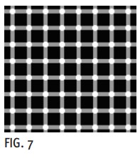
When looking at this illusion, there are gray spots that appear at the intersections of the white lines. These spots disappear as soon as you look at them
directly. Why? The answer has to do with how the retina converts visual stimuli into electrical impulses. According to Davies (2002), the darker
spots originate from what is known as lateral inhibition processing. Lateral inhibition is an important feature in the visual system, it enhances the
representation of contrast by improving the perception of edges (Kramer & Davenport, 2015). The Kanizsa Triangle (Fig. 8) was created by
Italian artist and psychologist Gaetano Kanizsa in 1955. This illusion is credited with making us realize how our visual system works by utilizing mechanisms of contour,
completion and fillingin, in addition to creating an illusion of depth. The Kanizsa Triangle is an essential part of the Gestalt
principles of organization (Wang et al., 2012).

The Muller-Lyer Illusion (Fig. 9) was created by German psychologist Franz Carl MullerLyer in 1889. A simple geometrical illusion of
size where the perceived length of the line in question is decreased by placing arrowheads at each end of the line and increased when placing arrow
tails (Zeman et al., 2013). The line on the right is perceived to be shorter than the one on the left when they are actually of
equal length. This is a distortion illusion.

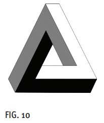
The Penrose Triangle (Fig. 10) was created by Oscar Reutersvard, known as the father of the impossible object, in 1934
and popularized by Lionel Penrose, a British psychiatrist and mathematician in 1958. This illusion is considered a paradox
or impossible objects illusion. The shape of the triangle is physically impossible to construct in the real world as it
would violate the rules of Euclidean geometry (Donaldson & Macpherson, 2017).
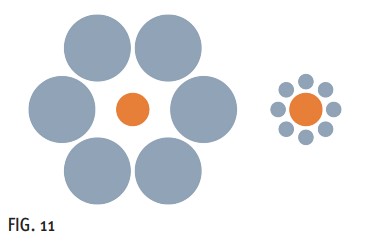
The Ebbinghaus Illusion (Fig.11), also known as Titchener’s circles, was discovered by German psychologist Herman Ebbinghaus in the 1890s.
This is a distortion illusion consisting of two inner circles of the same size. One of the circles is surrounded
by a group of larger circles, while a group of smaller circles surrounds the other. This context of differing-sized circles causes people to perceive the inner
circle surrounded by the smaller circles to be larger (Thomson & Macpherson, 2017).
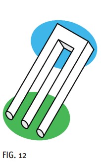
The Impossible Trident (Fig. 12) was created by D.H. Schuster, an American psychologist, in 1963.
This type of illusion is classified under paradox illusions depicting an impossible object that cannot exist. The impossible object, however, does not cease to
exist because the mind is modular, and the modules that form the visual system are “cognitively impenetrable” to the degree that the inner workings and outputs cannot be influenced
by conscious awareness (Donaldson, 2017).
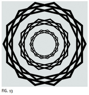
The Scintillating Starburst (Fig. 13) is a new class of illusion developed by Michael Karlovich, a visual artist and Pascal Wallisch, a psychology researcher.
The scintillating starburst is composed of concentric star polygons, and viewers see bright burst of rays emanating from its center.
This illusion illustrates how the brain connects the dots between certain points on the wreath, illustrating the subjective nature of reality in what we see and highlighting the
constructive nature of perception (Karlovich & Wallisch, 2021).
CONCLUSION
Even though the general population enjoys the entertainment value of optical illusions, they play a significant role in furthering our understanding of the process behind visual perception and our brains’ contribution to sight. Our brain constantly adds information to what our eyes report, filling in the blanks when needed to arrive at a “coherent” result. Optical illusions, however, find an open door to this “organized” process when there is a dissociation between the physical reality and the subjective perception of what we are observing. For those interested in this topic, I highly recommend reading The Oxford Compendium of Visual Illusions (9780199794607 Medicine & Health Science Books).